Scalable Imaging Workflows Advance Cancer Tissue Profiling
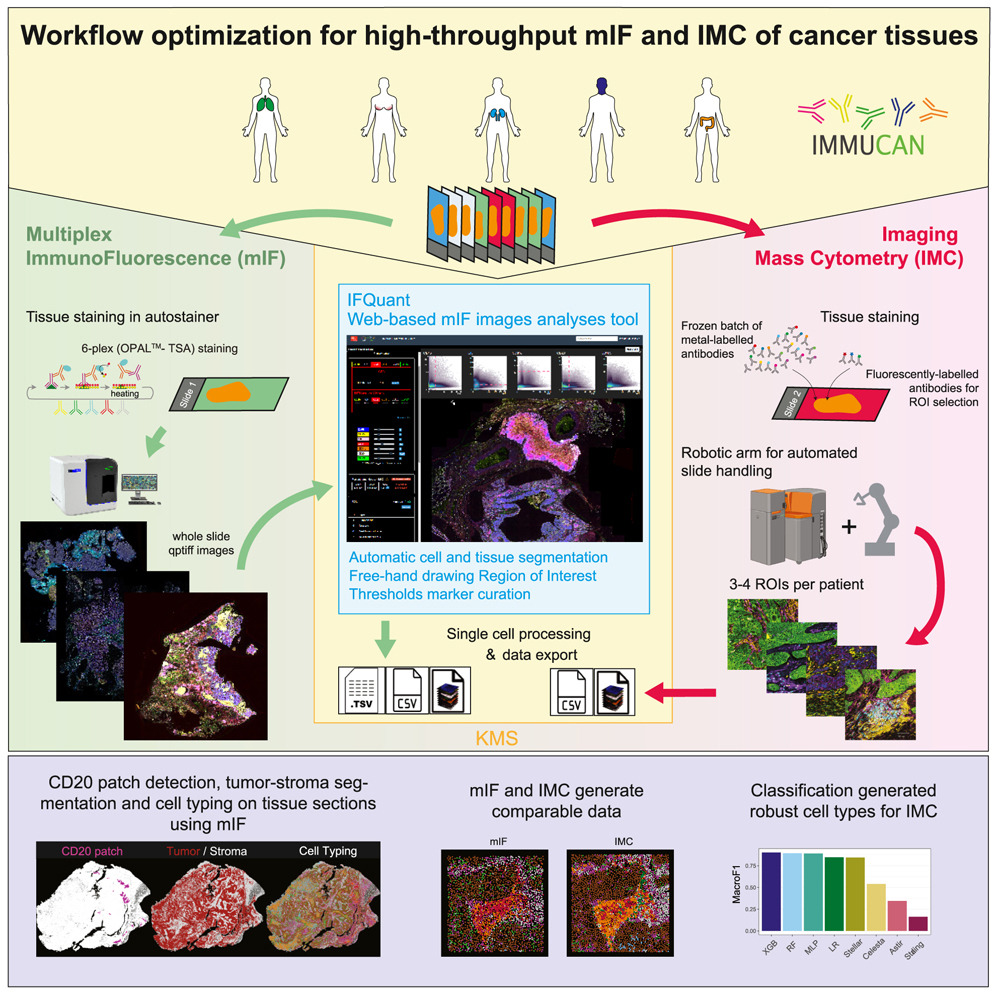
Researchers from the DQBM’s Bodenmiller Lab, in collaboration with partners across the IMMUcan consortium, have published a comprehensive methodology for high-throughput cancer tissue imaging in Cell Reports Methods. The study introduces two validated pipelines for multiplexed immunofluorescence (mIF) and imaging mass cytometry (IMC), applied to over 10,000 samples from more than 2,500 patients.
Key innovations include IFQuant, an open-source mIF analysis tool integrated with laboratory information systems, and a tree-based machine learning approach for robust IMC cell-type classification. The study reports high consistency between mIF and IMC data, ensuring reproducibility in immune landscape characterization across cancer types.
This multi-year effort marks a significant milestone in standardizing spatial single-cell analysis at scale, supporting translational oncology and biomarker discovery.
DOI: 10.1016/j.crmeth.2025.101170